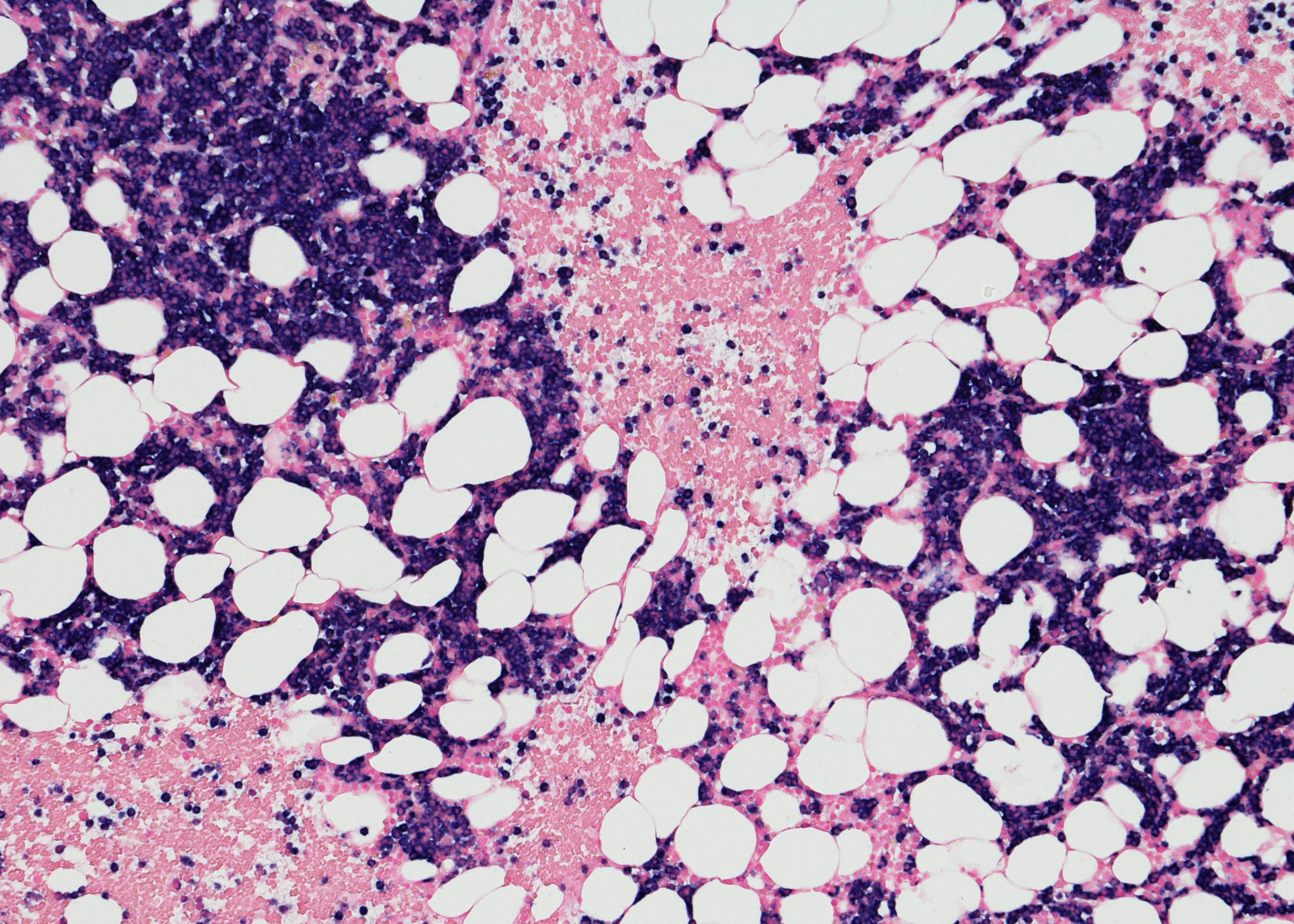
Multiple Myeloma.jpg

Multiple Myeloma – Symptoms, Causes & Treatment

Multiple myeloma is a type of cancer that develops in the bone marrow or the soft tissue inside your bones. It’s a malignant (cancerous) condition that occurs when abnormal plasma cells grow and invade other tissues, including the bone marrow. The abnormal cells produce large amounts of immunoglobulin, which can lead to various symptoms.

What are the Symptoms of Multiple Myeloma?

The most common signs and symptoms of multiple myeloma are:

  • Bone pain
  • Fatigue or weakness
  • Shortness of breath
  • Fever and chills
  • Loss of appetite (not related to nausea)

What are the Causes & Risk Factors of Multiple Myeloma?

There is no known cause of multiple myeloma. It is thought to be caused by a combination of factors, such as genetic predisposition, environmental exposure, and hormones, but several factors may increase your risk. These include:

  • Age – Multiple myeloma is most common in people over age 50.
  • Infection with Human T-cell Lymphotropic Virus Type 1 (HTLV-1) – A virus that can cause leukemia and other blood disorders; it’s found mostly in tropical areas.
  • Family History – People who have a family history of multiple myeloma are more likely to develop it themselves.
  • Exposure to radiation or certain chemicals

How is Multiple Myeloma Diagnosed?

A blood test, such as a complete blood count (CBC), can determine if your body has increased monoclonal immunoglobulin (Ig) G protein-specific antibodies. These antibodies are produced by the plasma cells that have been transformed into malignant plasma cells in multiple myeloma.

If the CBC determines that you have an increase in these proteins, your doctor will likely perform other tests. You may have a bone marrow aspiration and biopsy, which involves inserting a needle into your hip bone to remove some bone marrow for testing. This can help confirm the presence of cancerous plasma cells and determine whether they are located in areas where they shouldn’t be—such as the liver or kidneys.

Another common test for multiple myeloma is magnetic resonance imaging (MRI). This test creates images of your bones and soft tissues by sending radio waves through them so that they can be seen on a computer screen. An MRI can help detect tumors throughout your body and determine their size and location so that doctors can provide treatment plans accordingly.

How is it Treated?

Multiple myeloma can be treated with chemotherapy, targeted therapy, radiation therapy, and stem cell transplantation. Doctors will choose from these treatment options based on the patient’s medical history and type of multiple myeloma.

Chemotherapy is used to destroy cancer cells before they spread or return after treatment has ended. It does this by damaging or killing rapidly dividing cells that grow into tumors.

Targeted therapy uses drugs that target specific molecules involved in cancer growth or spread. These drugs may help stop cancer cells from growing or spreading so that they become less harmful to patients. Radiation therapy uses high-energy rays to destroy cancer cells.

Radiation may be used alone or combined with other treatments like chemotherapy or targeted therapy as part of your overall treatment plan for multiple myeloma.

Stem cell transplantation involves replacing some of your immune system’s stem cells with those from another person who has healthy stem cells but no history of cancer. Stem cell transplants are generally performed only when other treatments have not worked well enough.

Hematologists and Bone Marrow Transplantation experts at Burjeel Medical City, Abu Dhabi, treat multiple myeloma and other malignant blood diseases. They are also experts in bone marrow transplantation, stem cell transplantation, immunotherapy, and finding the best treatment for your condition.

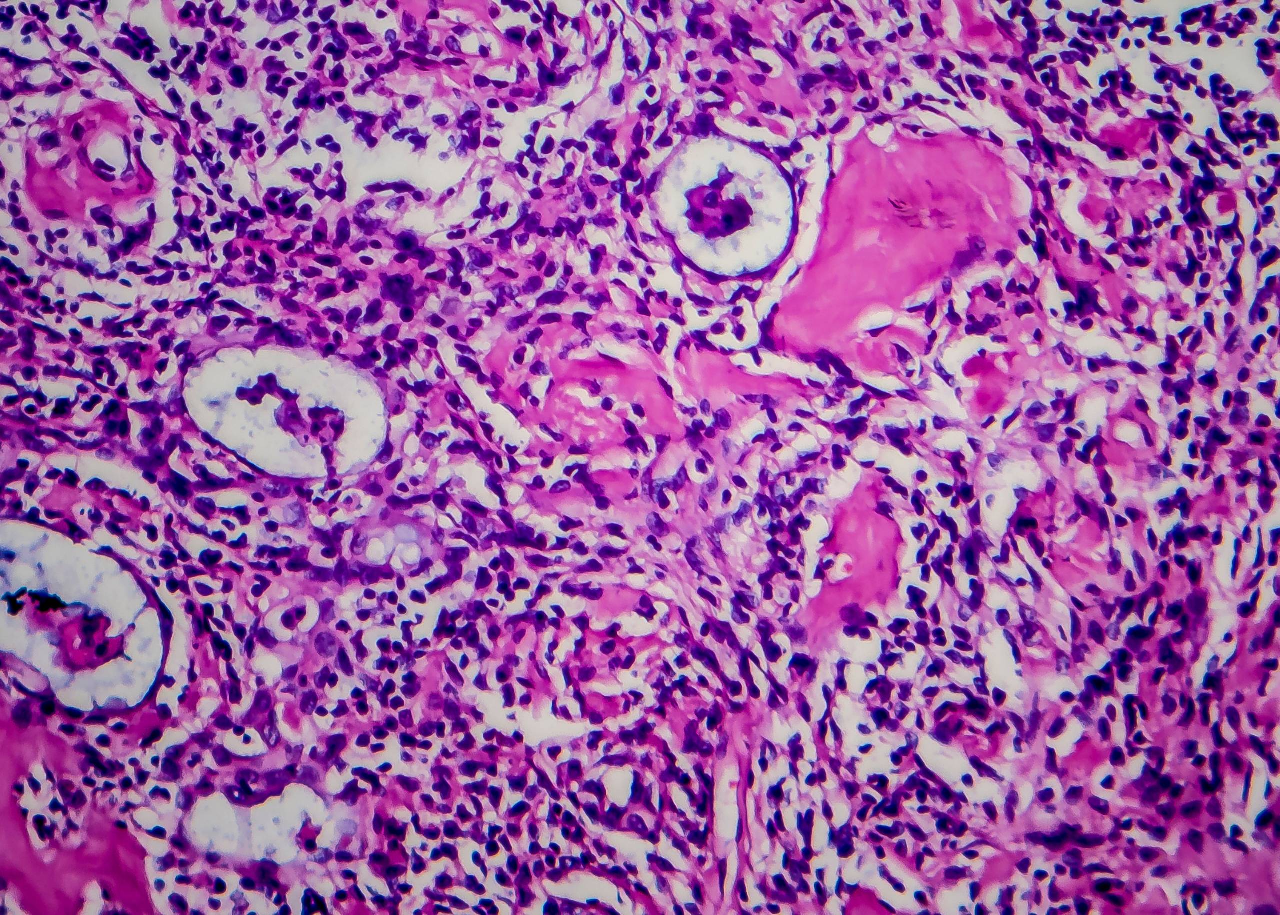
Myelodysplastic Syndromes.jpg

Myelodysplastic Syndromes – Symptoms, Causes & Treatment

Myelodysplastic syndromes (MDS) are a group of conditions that affect the blood and bone marrow. They are characterized by abnormal production of blood cells (myeloid cells), which can decrease the number or function of red blood cells, white blood cells, and platelets. MDS usually develops slowly over time and may progress to acute myeloid leukemia (AML).

What are the Symptoms of Myelodysplastic Syndromes?

The symptoms of MDS can vary from person to person and may not be present in all patients with this condition. Some of the most common symptoms include:

  • Feeling tired or short of breath
  • Feeling weak or dizzy
  • Bleeding or bruising more easily than normal
  • Pain in your bones or joints
  • Feeling itchy or tingling sensations in your arms or legs
  • Feeling like you have a fever

What are the Types of Myelodysplastic Syndromes?

  • Acute Myeloid Leukemia, or AML, is the most common type of myelodysplastic syndrome. It occurs when the bone marrow produces too many immature white blood cells, which can cause infections, anemia, and bleeding disorders. The leukemia cells can also spread to other parts of your body.
  • Chronic Myelogenous Leukemia (CML)—This type of leukemia affects older adults but is not as common as AML. CML causes abnormal growth of white blood cells in the bone marrow. If untreated for a long time, it can progress into acute leukemia or another form of cancer called acute promyelocytic leukemia (APL).
  • Juvenile Myelomonocytic Leukemia (JMML)—This form affects children under age 20. It is less likely than other types of myelodysplastic syndrome to be inherited from your parents’ genes because it usually appears without warning during childhood or adolescence without any family history of JMML or other medical conditions.

What is the Cause of Myelodysplastic Syndromes?

The cause of MDS is unknown. It can develop in people who have had previous radiation or chemotherapy treatment. Other risk factors include aging, having a family history of MDS, being exposed to certain chemicals or drugs, and having certain genetic conditions.

How is it Diagnosed?

Myelodysplastic Syndromes can be diagnosed by a number of different methods, including:

Blood tests: These are used to measure red blood cell counts and the number of platelets in the body. They may also check for white blood cells and other markers that can signal disease.

Bone marrow biopsy: A small amount of bone marrow is extracted from your hip bone, which is then examined under a microscope. If your bone marrow is unhealthy, it will show signs of myelodysplastic syndrome.

Lumbar puncture: This test involves inserting a needle into your spine to collect cerebrospinal fluid, which surrounds the brain and spinal cord. The fluid can be tested for signs of leukemia or other cancers in addition to myelodysplastic syndromes.

How is it Treated?

Myelodysplastic syndromes are treated with a combination of chemotherapy, radiation therapy, and bone marrow transplantation. The goal is to slow down or stop the progression of this disease.

Chemotherapy is often used to treat myelodysplastic syndromes. This type of treatment involves using certain drugs to kill cancer cells and shrink tumors. These drugs are typically taken by mouth or injected into a vein (IV). Some chemotherapy drugs can be given through an IV in your arm or hand before you go home from the hospital or doctor’s office after surgery to remove your spleen. Radiation therapy may also be used to treat this condition.

Radiation therapy is a cancer treatment that uses high-energy x-rays or other types of radiation to destroy cancer cells and keep them from growing back (recurring).

If these treatments don’t work, you may need a bone marrow transplantation. A bone marrow transplantation replaces damaged bone marrow with healthy bone marrow that contains normal blood cells.

Hematologists and Bone Marrow Transplantation experts at Burjeel Medical City, Abu Dhabi, treat Myelodysplastic Syndromes with the latest medical advancements

Hodgkin Lymphoma.jpg

Hodgkin Lymphoma – Symptoms, Causes & Treatment

Hodgkin lymphoma is a cancer of the lymphatic system. The lymphatic system is a network of glands, nodes, and channels producing lymph fluid. The lymphatic system helps the body fight infection by removing bacteria and other foreign substances from the blood. It also helps fight cancers and tumors by making special proteins called antibodies.

It affects the lymph nodes and other parts of the immune system. It usually progresses slowly but can be life-threatening if it spreads to other organs like the lungs or bone marrow.

Hodgkin lymphoma is named for description by Thomas Hodgkin and his colleagues at Guy’s Hospital in London in 1832.

Types of Hodgkin Lymphoma

There are four main types: 

Nodular Sclerosing Hodgkin Lymphoma (NSHL) is a rare form of Hodgkin lymphoma with a distinctive appearance. It is characterized by large, solid tumors that are sessile and have no capsule. These nodules are surrounded by a thin rim of reactive cells, which can appear as a pale halo surrounding the nodule on CT scans. The lesions may be solitary or multiple and can occur anywhere in the body.

Nodular Lymphocyte Predominant Hodgkin Lymphoma (NLPHL) is a rare, aggressive form of non-Hodgkin lymphoma. A distinctive pattern of histology and molecular alterations characterizes it. Affected individuals are typically younger than those who have other types, and the disease tends to be more aggressive in its early stages.

Lymphocyte Rich Classical Hodgkin Lymphoma (LRCH) is a type of Hodgkin lymphoma that is characterized by an abundance of lymphocytes (white blood cells), which are the main component of the immune system. It is also known as “nodular sclerosis classical Hodgkin lymphoma” or “nodular lymphocyte predominant Hodgkin lymphoma.” The term “nodular” refers to the characteristic tumor nodules appearing in Hodgkin lymphoma. In contrast, there are fewer or no nodules in most other forms. Two types of LRCH exist type 1, which has a more favorable prognosis than type 2 and type 2, which has a worse prognosis than type 1.

Mixed Cellularity Classical Hodgkin Lymphoma (MCCL) is a form that has cells with both Reed-Sternberg and non-Reed-Sternberg features. It is more common in older adults than other types.

Symptoms of Hodgkin Lymphoma

The most common symptoms are:

  • Painless swelling of the neck, underarms, or groin
  • A fever that lasts for more than 3 days
  • Night sweats
  • Weight loss for no known reason

Causes of Hodgkin Lymphoma

The cause of this disease is unknown, but research has shown that it could be linked to genetics and lifestyle factors. In particular, a person’s risk of developing the condition increases if they have a family history of the illness or if they have been exposed to certain environmental factors like radiation or chemicals.

Risk Factors of Hodgkin Lymphoma

The risk factors include,

Age. Hodgkin lymphoma is most common in people between the ages of 15 and 30, with the highest rates occurring among those between 20 and 24 years old. While it can occur at any age, it is rare in children younger than 10.

Gender. Males are more likely to develop the condition than females. However, it is important to note that this difference may be due in part to the fact that men are more likely to seek medical care when they experience symptoms.

Ethnicity. Caucasians are more likely to develop the condition than Asians or African Americans.

Family History. People who have a parent or sibling with Hodgkin lymphoma have an increased risk of developing it themselves. In addition, there is evidence that suggests having multiple family members with the condition can increase your risk by two times compared with someone who does not have this family history.

Diagnosis

Tests and procedures used to diagnose the condition include the following: 

Physical Exam: The doctor will examine you, checking for swollen lymph nodes in your neck, underarm area, or groin. They’ll also check your skin for signs of infection and look at your body for any other abnormalities.

Blood Tests: A blood test can confirm the presence of Hodgkin’s lymphoma by looking for increased levels of a protein called LDH (lactate dehydrogenase). Chest X-ray: An X-ray can help determine whether you have any enlarged lymph nodes in your lungs or chest wall. It may also show signs of lung damage from cancer treatment.

CT Scan: A computed tomography (CT) scan uses X-rays to make detailed pictures of areas inside the body, including the lymph nodes. This test can determine if there are enlarged lymph nodes and whether they contain cancer cells. 

MRI: Magnetic resonance imaging (MRI) uses a strong magnetic field and radio waves to make detailed pictures of areas inside the body without using X-rays. It can help doctors see tumors that are difficult for CT scans to detect. 

Lymph Node Biopsy: The doctor will use a needle to remove several small tissue samples from your lymph nodes for testing in the lab. In some cases, this may be done under sedation or general anesthesia so the procedure doesn’t cause pain or discomfort.

Treatment

Treatment involves chemotherapy, radiation therapy, and stem cell transplantation.

Chemotherapy is used to treat both stages 1 and 2 of Hodgkin lymphoma. The goal is to destroy cancer cells with drugs against fast-growing cells. Chemotherapy may be combined with radiation therapy or surgery to treat the condition.

Radiation therapy uses high-energy rays or particles to kill cancer cells. Radiation therapy may be used alone or in combination with chemotherapy to treat the condition.

Stem cell transplantation is used if your doctor thinks your disease has come back after treatment (recalled) or if you have advanced stage 3 or 4 Hodgkin lymphoma. Stem cells are immature blood cells that can become any blood cell in your body when they mature. Stem cell transplants replace your unhealthy bone marrow with healthy donor bone marrow.

Hematologists and Bone Marrow Transplanation experts at Burjeel Medical City, Abu Dhabi, treat Hodgkin lymphoma and other blood disorders.

Liver Cancer.jpg

Liver Cancer – Symptoms, Causes & Treatment

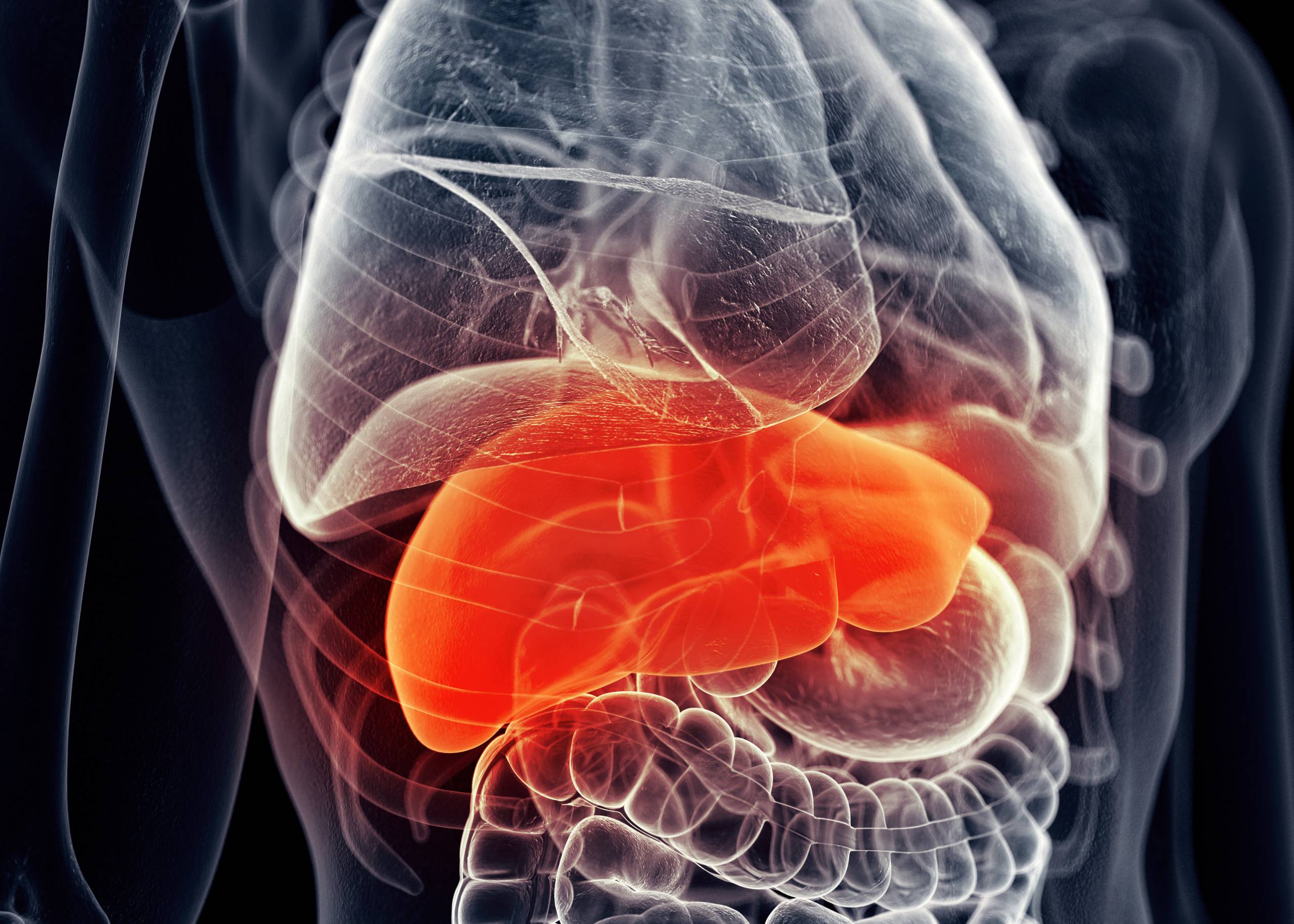
Liver cancer is a disease in which malignant (cancerous) cells form in the liver. The word “liver” can also refer to other abdominal organs that perform similar functions, including the gallbladder, stomach, and pancreas. Liver cancer may also be called hepatocellular carcinoma (HCC), which means “cancer of the liver.” The liver is a large gland in the upper right of the abdomen. It performs many functions, including detoxifying blood, producing bile to help digest fats, and storing vitamins A and D. The liver also helps regulate blood sugar (glucose) levels by converting excess glucose into glycogen for storage in muscles or fat cells.

Symptoms of Liver Cancer

The symptoms depend on the stage of cancer. In general, symptoms include:

  • Vomiting (nausea) or feeling like you can’t keep anything down
  • Jaundice (yellowing of the skin, mucous membranes, and whites of the eyes)
  • Loss of appetite or weight loss -Fatigue or weakness

Causes of Liver Cancer

A number of different factors causes liver cancer. Some of these include:

  • Hepatitis B and C infections
  • Liver diseases, such as cirrhosis or hepatitis
  • Alcohol abuse
  • Cigarette smoking
  • Obesity

Hepatitis B virus (HBV) infection accounts for about one-third of all cases of HCC worldwide and about 70% in Asia. Other causes include hepatitis C virus (HCV), cirrhosis due to alcohol abuse, and nonalcoholic fatty liver disease (NAFLD).

Diagnosis

It is diagnosed with imaging, blood tests, and biopsies. Imaging can be done with ultrasound, CT, MRI, or PET scans.

  • An ultrasound uses high-frequency sound waves to produce an image of the liver on a monitor.
  • CT scans use X-rays to create three-dimensional images of internal structures. MRIs use radio waves and magnetic fields to generate images of the body’s internal structures.
  • PET scans use a small amount of radioactive material that attaches itself to cancer cells so the scanner can detect them.
  • Blood tests check for certain proteins in the blood that may indicate cancerous tissue growth. These proteins include AFP (alpha fetoprotein), CA 19–9 (carbohydrate antigen 19–9), and CA 72–4 (carbohydrate antigen 72–4).
  • Biopsies are performed on the liver tissue to confirm whether or not cancer is present.

Treatment

Liver cancer is a serious disease that can be fatal if not treated. The main goal of treatment is to cure cancer, but this may not always be possible. Treatment options depend on the type and stage of your liver cancer.

The main treatment options for early-stage liver cancer include surgery and chemotherapy.

Surgery may include a partial hepatectomy (removing part of your liver) or a liver transplant.

More advanced tumors may require chemotherapy or radiation therapy.

Liver transplantation is an option for patients who have a liver tumor that cannot be removed by surgery or when their tumors have spread to other parts of their body. The surgery involves removing the patient’s diseased liver and replacing it with a healthy one from another person or donor organ.

Experts in the Department of Hepatopancreatobiliary Surgery and Transplantation at Burjeel Medical City (BMC), Abu Dhabi, treats patients suffering from liver, pancreas, and biliary system ailment.

Liver Cancer & Transplant Expert

Dr. Rehan Saif Web

Dr. Rehan Saif

Director of Transplant Surgery for Burjeel Abdominal Multi-Organ Transplant Program, Clinical Lead HPB Surgery, Consultant General Surgery

Lung Cancer.jpg

Lung Cancer – Symptoms, Causes & Treatment

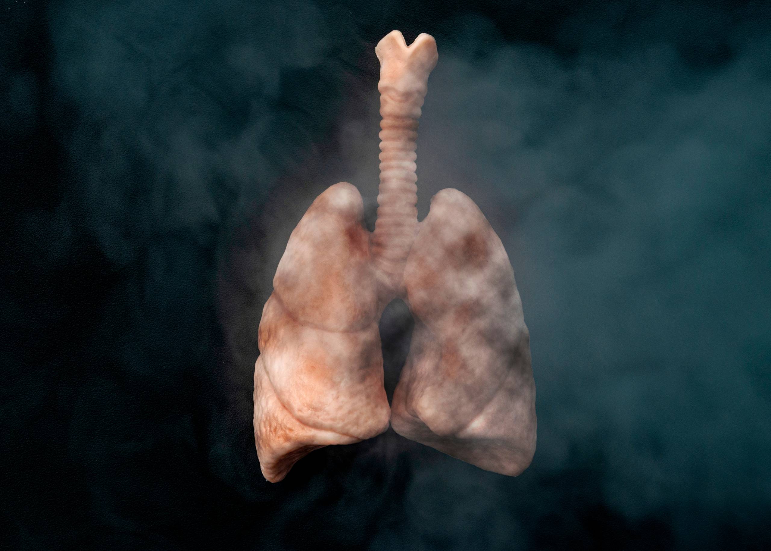
Lung cancer occurs when abnormal cells in the lungs divide uncontrollably and form tumors. These tumors can block airflow to the lungs, making breathing difficult for people with the condition. It most often develops in the tissues of the bronchi, which are the airways that carry oxygen-rich air from your mouth to your lungs. It can also develop in other parts of the lung, including the pleura (the lining between your lungs and chest wall), alveoli (small sacs where oxygen is exchanged for carbon dioxide), lymph nodes within the chest cavity, or space in between the lungs.

Symptoms of Lung Cancer

The symptoms are similar to those of other illnesses, so it can be difficult to tell if you have the condition. The most common symptoms include:

  • Changes in your cough
  • Chest pain, which may be worse when you breathe deeply
  • Shortness of breath, which may be worse when you lie down
  • Blood in your phlegm (spit) or coughing up blood

Causes of Lung Cancer

It is the second most common type of cancer in the world, and it is the leading cause of cancer-related death. It’s also one of the most preventable cancers: quitting smoking lowers your risk by 50%. But what are some other causes of lung cancer? Here are some of the more common ones:

  • Exposure to secondhand smoke
  • Radon gas exposure
  • Air pollution (including particulate matter)
  • Diesel exhaust fumes

Diagnosis

It is diagnosed by a combination of physical examination, imaging, and biopsy. Imaging tests include chest X-rays, CT scans, MRI scans, PET scans, and ultrasounds. These tests will show whether or not you have any tumors in your lungs. Finally, a biopsy is performed to confirm the presence of lung cancer. A sample of tissue is taken from the tumor under local anesthesia.

Treatment

Lung cancer is the leading cause of cancer-related deaths worldwide. The five-year survival rate for lung cancer patients is only 16 percent. There are several types of treatment, including surgery, chemotherapy, radiation therapy, and targeted therapy.

Surgery is used to remove tumors in the lung or nearby lymph nodes.

Chemotherapy uses drugs to kill cancer cells or stop them from growing.

Radiation therapy uses high-energy rays to destroy cancer cells. Targeted therapy uses drugs that block the growth and spread of cancer cells. The best treatment depends on your age, health, and type of lung cancer. If you or your loved ones have lung cancer, talk with your doctor about the best treatment options for you.

Burjeel Medical City, Abu Dhabi, is one of the leading cancer treatment centers in the Middle East. The hospital offers a wide range of treatment options for lung cancer patients, including surgery and radiation therapy. The center also offers advanced diagnostic technologies, such as CT and MRI scans, to identify the stage of your cancer.

20612 BURJEEL BREAST CANCER WEBSITE BANNER WEB BANNER 2 1.jpg

Detecting Early Symptoms of Breast Cancer: A Guide for Women

Breast cancer is one of the most common cancers affecting women globally. The good news is that when detected early, it can be treated successfully. Early detection plays a vital role in improving survival rates, which is why it’s important for every woman to know the signs and symptoms of breast cancer and stay vigilant about her breast health.

Why Early Detection Matters

Breast cancer doesn’t usually cause pain or noticeable symptoms in its earliest stages, making regular screening and awareness crucial. When caught early, breast cancer treatment is often more effective and less invasive, with higher survival rates. That’s why self-awareness and routine medical checkups are key to catching breast cancer before it spreads.

Early Symptoms of Breast Cancer to Watch For

Breast cancer can present itself in a variety of ways. It’s important to know your body and recognize changes. Here are some common signs that may indicate early breast cancer:

1. Lump or Mass in the Breast:
One of the most common symptoms of breast cancer is the presence of a lump or mass in the breast. This mass is often hard and painless, but in some cases, it can feel tender or soft. Not all lumps are cancerous, but if you find one, it’s critical to consult your doctor for further evaluation.

2. Changes in Breast Size or Shape:
Noticeable changes in the size or shape of one breast compared to the other can be an early indicator of breast cancer. Sudden swelling, shrinkage, or asymmetry should not be ignored.

3. Skin Changes on the Breast:
Breast cancer can sometimes cause changes to the skin covering the breast, including:

  • Dimpling or a texture resembling an orange peel.
  • Redness or a rash that doesn’t go away.
  • Thickening or swelling in the breast.
    These changes may be subtle, but they’re important to discuss with your healthcare provider.

4. Nipple Changes:
Nipple changes are another potential warning sign of breast cancer. Keep an eye out for:

  • Inverted or retracted nipples (when the nipple turns inward).
  • Discharge from the nipple, especially if it’s bloody or occurs without squeezing.
  • Scaling, peeling, or flaking skin around the nipple.
    While some nipple changes can be linked to hormonal shifts, they could also signal underlying issues like breast cancer.

5. Unexplained Breast Pain:
While breast pain is more commonly associated with hormonal changes, persistent or unusual pain in one area of the breast that doesn’t go away with your menstrual cycle could be a sign of something more serious.

6. Lumps in the Armpit:
Breast cancer can spread to nearby lymph nodes, including those in the armpit area. If you notice any unusual swelling or lumps in your armpit, it’s important to bring this to your doctor’s attention.

How to Detect Breast Cancer Early

There are several steps you can take to ensure breast cancer is detected at its earliest and most treatable stage:

1. Perform Regular Breast Self-Exams
Knowing what’s normal for your breasts can help you detect changes early. A breast self-exam should be done monthly, ideally a few days after your period ends. Here’s how to perform a self-exam:

  • In front of a mirror, inspect your breasts with your arms at your sides and then raised overhead. Look for changes in size, shape, and skin texture.
  • Lying down, use the pads of your fingers to feel your breasts in circular motions, checking the entire breast, armpit, and collarbone area.
  • In the shower, feel for any lumps or changes with soapy hands, which can help detect subtle differences.
    If you notice any changes, make an appointment with your doctor for further evaluation.

2. Get Regular Mammograms
Mammograms are the most reliable method for detecting breast cancer in its early stages. Most women should start having mammograms at age 40, but if you have a family history of breast cancer or other risk factors, your doctor may recommend starting earlier.
Mammograms can detect tumors before they’re large enough to feel and can identify abnormal areas that need further investigation. Make sure to schedule a mammogram as part of your regular health check-up.

3. Clinical Breast Exams
In addition to self-exams and mammograms, your OB-GYN or primary care physician should perform a clinical breast exam during your annual check-up. This is an important opportunity to discuss any changes you’ve noticed and receive a professional evaluation.

4. Genetic Testing for High-Risk Women
Women with a family history of breast cancer may benefit from genetic testing to determine if they carry BRCA1 or BRCA2 gene mutations, which increase the risk of breast and ovarian cancers. If you’re identified as high-risk, your doctor may recommend more frequent screenings or preventive measures.

Reducing Your Risk of Breast Cancer

While some risk factors for breast cancer, such as age and family history, are out of your control, there are several lifestyle choices you can make to reduce your risk:

  • Maintain a healthy weight: Obesity, especially after menopause, has been linked to an increased risk of breast cancer.
  • Exercise regularly: Staying active helps maintain a healthy weight and lowers your risk.
  • Limit alcohol consumption: Excessive alcohol intake is associated with a higher risk of breast cancer.
  • Breastfeed: Breastfeeding has been shown to lower the risk of developing breast cancer, especially in women who breastfeed for over a year.
  • Avoid smoking: Smoking has been linked to many types of cancer, including breast cancer.

Conclusion: Stay Vigilant and Informed

Early detection of breast cancer is critical for successful treatment, and staying informed about the early warning signs can make all the difference. Be proactive about your breast health—perform regular self-exams, schedule routine mammograms, and maintain a healthy lifestyle.
At Burjeel Hospital, our team of experts is dedicated to providing comprehensive care for women. If you have concerns about your breast health or want to schedule a screening, our experienced doctors are here to guide you through the process with compassion and expertise.

22050 BHAD Blog Creative The Laparoscopic Revolution Why Less Invasive Surgery Is The Future Website 1.jpg

The Laparoscopic Revolution: Why Less Invasive Surgery is the Future 

Surgical advancements have transformed healthcare, offering patients safer, less painful, and faster recovery options. One of the most significant innovations is laparoscopic surgery—a minimally invasive technique that is reshaping the future of surgical care. More recently, robotic-assisted surgery has taken laparoscopic procedures to the next level, enhancing precision and improving patient outcomes.

In this blog, we’ll explore why laparoscopic surgery is gaining momentum, its impact on various health conditions, and how it benefits patients by reducing risks and improving recovery times. We’ll also discuss its critical role in colorectal surgery, where minimally invasive techniques are revolutionizing treatments for conditions like colorectal cancer, inflammatory bowel disease (IBD), and diverticulitis.

What is Laparoscopic Surgery?

Laparoscopic surgery, also known as minimally invasive surgery, uses small incisions and specialized instruments to perform procedures with minimal disruption to the body. Unlike traditional open surgeries that require large incisions, this technique allows surgeons to operate with precision using a tiny camera (laparoscope) and specialized tools. Recently, robotic-assisted laparoscopic surgery has been introduced, allowing for even greater accuracy, flexibility, and control during procedures.

The Benefits of Laparoscopic Surgery

  • Reduced Pain and Discomfort – Smaller incisions mean less trauma to surrounding tissues, leading to significantly reduced post-operative pain.
  • Faster Recovery – Many laparoscopic procedures allow patients to return to their normal activities within days rather than weeks.
  • Lower Risk of Infection – Smaller incisions reduce exposure to external contaminants, minimizing the risk of post-surgical infections.
  • Minimal Scarring – Because the incisions are small, scarring is significantly less noticeable compared to traditional surgery.
  • Shorter Hospital Stays – Many laparoscopic procedures can be done on an outpatient basis or require only a short hospital stay, lowering healthcare costs and improving patient comfort.
  • Enhanced Precision with Robotics – Robotic technology used in laparoscopic procedures provides surgeons with an enhanced range of motion, 3D visualization, and more accurate movements, improving outcomes and reducing complications.

Laparoscopic Surgery and Colorectal Conditions

Laparoscopic surgery has transformed the treatment of colorectal diseases, offering a minimally invasive approach to conditions that previously required extensive open surgery.

  1. Laparoscopic Surgery for Colorectal Cancer

Laparoscopic and robotic-assisted techniques have become the standard of care for many colorectal cancer patients. These procedures allow for precise removal of cancerous tissue while preserving as much healthy bowel as possible, leading to faster recovery and fewer complications.

  1. Treatment for Inflammatory Bowel Disease (IBD)

For patients with Crohn’s disease or ulcerative colitis requiring surgery, laparoscopic procedures can reduce post-operative pain and shorten recovery times. Minimally invasive bowel resections or stricturoplasties help improve quality of life with fewer complications.

  1. Managing Diverticulitis with Minimally Invasive Surgery

Severe cases of diverticulitis may require surgery, and laparoscopic approaches allow for safer bowel resections with less pain and faster healing. This is especially beneficial for patients with recurrent or complicated diverticulitis.

Key Procedures Performed Using Laparoscopy

Laparoscopic surgery is revolutionizing treatments across multiple specialties, particularly in colorectal and abdominal surgery:

  • Colectomy (Colon Resection) – A minimally invasive technique to remove diseased parts of the colon in conditions like colorectal cancer, Crohn’s disease, and diverticulitis.
  • Rectal Surgery – Laparoscopic or robotic-assisted rectal resections improve precision and reduce complications in rectal cancer and other diseases.
  • Gallbladder Removal (Cholecystectomy) – A common laparoscopic procedure to remove the gallbladder with minimal recovery time.
  • Hernia Repair – Minimally invasive hernia surgeries reduce pain and accelerate healing.
  • Bariatric Surgery – Weight-loss procedures help in reducing risk factors for obesity-related conditions like diabetes and high cholesterol levels.
  • Appendectomy – Emergency laparoscopic appendectomy shortens hospital stays and speeds up recovery.
  • Gynecological ProceduresMinimally invasive hysterectomy, ovarian cyst removal, and fibroid treatments.

In many of these cases, robotic systems further enhance the surgeon’s ability to navigate and control surgical instruments with a high degree of precision.

Diet and Lifestyle After Laparoscopic Surgery

Recovery from laparoscopic procedures is generally smoother, but certain dietary and lifestyle changes can enhance healing:

  • Nutrient-rich foods for diabetics can aid in stabilizing blood sugar post-surgery.
  • Regular exercise supports faster recovery by improving circulation and preventing complications.
  • Monitoring vital signs, such as cholesterol levels and blood pressure, ensures long-term success.
  • Preventive strategies, including balanced nutrition and routine check-ups, help sustain health benefits.

Frequently Asked Questions (FAQs)

Is laparoscopic surgery safe for people with diabetes? 

Yes, laparoscopic surgery is generally safe for diabetic patients. In fact, some procedures, such as bariatric surgery, can help improve insulin sensitivity and blood sugar control. However, blood sugar levels must be monitored before and after surgery.

How long is the recovery time after laparoscopic surgery? 

Recovery time varies depending on the procedure, but most patients can return to daily activities within a few days to a week. Strenuous activities should be avoided for several weeks.

Can laparoscopic surgery help with vascular health? 

Yes, certain laparoscopic procedures can improve vascular function, especially in conditions like peripheral artery disease (PAD) or varicose veins, by enhancing blood flow with minimal trauma.

What are the long-term benefits of minimally invasive surgery? 

The long-term benefits include faster recovery, minimal scarring, lower risk of infection, and improved overall health outcomes, especially for patients with conditions like diabetes, hypertension, and obesity.

Are there any dietary restrictions after laparoscopic surgery? 

While there are no strict restrictions, patients are advised to consume nutrient-dense foods that support healing, such as lean proteins, vegetables, and whole grains. Those with diabetes should monitor their carbohydrate intake carefully.

Why Laparoscopy and Robotics is the Future of Surgery

With the rise of technological advancements and increasing demand for less invasive medical interventions, laparoscopic surgery is becoming the gold standard for various procedures. Robotic-assisted laparoscopic surgery is at the forefront of this shift, offering even more precision, flexibility, and faster recovery times. As the healthcare industry continues to evolve, minimally invasive techniques, powered by robotics, will play a vital role in ensuring faster, safer, and more efficient treatments for a wide range of conditions.

If you’re considering surgery, consult with a surgeon or a specialist to determine whether laparoscopic or robotic surgery is right for you. The future of surgery is here—offering better outcomes, faster recovery, and a path toward improved health.

Ready to explore your options? Schedule a consultation with our doctors at Burjeel hospital Abu-Dhabi today!

MRI Negative Cushing Disease  Advanced Neurosurgical Success Story.jpg

MRI-Negative Cushing Disease: Advanced Neurosurgical Success Story

When a 32-year-old man arrived at Burjeel Medical City seeking a second opinion for his mysterious symptoms, he had already been diagnosed with Cushing’s disease at another facility. However, his case would prove to be one of the most challenging endocrine disorders to diagnose and treat – a condition so rare that it affects only 1-2 people per million worldwide.

The Journey to Accurate Diagnosis

The patient’s health concerns began subtly with decreased libido, a symptom that might seem minor but was actually the first indicator of a serious hormonal imbalance. Medical evaluation revealed hypogonadotropic hypogonadism, a condition where the body doesn’t produce enough reproductive hormones.

As his condition progressed, more concerning symptoms emerged:

  • Significant weight gain with central obesity
  • Purple stretch marks (striae) appearing on his trunk
  • Development of “moon face” – a characteristic rounded facial appearance
  • Formation of a “buffalo hump” – fatty deposits between the shoulders

These physical changes, combined with his hormonal abnormalities, painted a clear picture of Cushing’s disease – a rare condition caused by excess cortisol production.

The Diagnostic Mystery: When Advanced Imaging Fails

Despite having classic symptoms of Cushing’s disease, the patient’s case presented an extraordinary challenge. Laboratory tests confirmed the diagnosis with elevated hormone levels:

  • 24-hour urine free cortisol: 371 mcg (significantly elevated)
  • Morning serum cortisol: 24.72 (above normal range)
  • ACTH levels: 64.3 (indicating pituitary origin)

However, both 1.5 Tesla and 3 Tesla MRI scans showed no visible pituitary tumor. This presented what medical experts call “MRI-Negative Cushing Disease” – a condition that represents approximately 25% of all Cushing’s disease cases and creates significant diagnostic and treatment challenges.

Advanced Diagnostic Techniques: The Role of Multidisciplinary Care

Recognizing the complexity of the case, Burjeel Medical City’s team implemented a comprehensive multidisciplinary approach involving:

Endocrine Tumor Board Consultation

The case was presented to a specialized tumor board comprising endocrinologists, neurosurgeons, interventional radiologists, and pathologists. This collaborative approach ensures that every aspect of the patient’s condition is carefully evaluated.

Inferior Petrosal Sinus Sampling (IPSS)

When standard imaging fails to locate a pituitary tumor, IPSS becomes the gold standard for tumor localization. This sophisticated procedure involves:

  • Catheter insertion into the inferior petrosal sinuses
  • Direct hormone sampling from blood vessels draining the pituitary gland
  • Precise tumor localization by comparing hormone levels from different sides

In this patient’s case, IPSS revealed significantly elevated ACTH levels in the left inferior petrosal sinus, confirming the tumor’s location on the left side of the pituitary gland.

Cutting-Edge Surgical Intervention: Minimally Invasive Endoscopic Approach

Dr. Mohamed A. Elzoghby, Consultant Neurosurgeon at Burjeel Medical City, led the surgical team in performing a minimally invasive endoscopic transsphenoidal resection. This advanced technique offers several advantages over traditional open surgery:

The Oldfield’s Technique

This specialized surgical approach involves:

  • Extracapsular dissection to identify the tumor’s pseudo-capsule
  • Precise tumor removal while preserving healthy pituitary tissue
  • Minimal tissue disruption compared to traditional methods

Endoscopic Advantages

  • Panoramic visualization of the surgical field
  • High magnification for precise tumor identification
  • Improved tumor detection even when invisible on MRI
  • Faster recovery compared to open procedures

The Expert Surgical Team

This complex case required the expertise of multiple specialists:

Remarkable Recovery and Patient Outcomes

The surgery was a complete success, with several positive indicators:

Immediate Surgical Success

  • Complete tumor removal achieved using microsurgical techniques
  • Preservation of normal pituitary function
  • No intraoperative complications

Biochemical Remission

Post-operatively, the patient’s cortisol levels dropped below 0.8 µg/dL, confirming complete remission of Cushing’s disease. This dramatic improvement indicated successful removal of the ACTH-secreting adenoma.

Rapid Recovery

  • Started on cortisol replacement therapy as a temporary measure
  • Discharged after only 3 days without complications
  • Excellent recovery at 6-week follow-up

Understanding the Challenges of MRI-Negative Cushing Disease

This case highlights several important aspects of rare endocrine disorders:

Diagnostic Complexity

  • 25% of Cushing’s cases are MRI-negative
  • Delayed diagnosis often occurs due to imaging limitations
  • Specialized testing like IPSS is crucial for proper localization

Treatment Challenges

  • Extensive pituitary exploration may be required during surgery
  • Advanced visualization techniques improve tumor identification
  • Multidisciplinary expertise is essential for optimal outcomes

Why Burjeel Medical City Excels in Complex Endocrine Surgery

This successful case demonstrates several key strengths of Burjeel’s approach:

Advanced Technology

  • High-resolution 3 Tesla MRI for detailed imaging
  • State-of-the-art endoscopic equipment for minimally invasive surgery
  • Intraoperative imaging capabilities for real-time guidance

Specialized Expertise

Multidisciplinary Care

Patient Testimonial: Excellence in Care

The patient expressed profound gratitude for his treatment experience:

“I would like to express my deepest gratitude to Dr. Mohamed A. Elzoghby and his incredible team for the exceptional care I received throughout my recent surgery. Dr. Elzoghby’s professionalism, precision, and calm confidence immediately put me at ease… What stood out to me most was not just his surgical expertise, but his genuine care and attentiveness.”

The Future of Rare Endocrine Disorder Treatment

This case represents the evolution of medical care for complex endocrine conditions:

Precision Medicine

  • Targeted diagnostic approaches for rare conditions
  • Minimally invasive surgical techniques reduce patient morbidity
  • Personalized treatment plans based on individual patient needs

Technology Integration

  • Advanced imaging continues to improve tumor detection
  • Robotic assistance may further enhance surgical precision
  • Real-time monitoring improves surgical outcomes

Conclusion: Hope for Patients with Rare Conditions

The successful treatment of MRI-negative Cushing disease at Burjeel Medical City demonstrates that even the most challenging medical conditions can be effectively managed with:

  • Specialized expertise in rare endocrine disorders
  • Advanced diagnostic techniques like IPSS
  • Minimally invasive surgical approaches
  • Comprehensive multidisciplinary care
  • State-of-the-art medical technology

For patients facing similar challenges, this case provides hope that expert medical teams can achieve excellent outcomes even in the most complex situations.

Osseointegration Procedure For Transtibial Amputees.png

Pioneering Osseointegration Surgery Transforms Life for Transtibial Amputee

Introduction

Burjeel Medical City continues to lead the way in advanced surgical interventions with a groundbreaking osseointegration procedure performed on a 57-year-old patient with a history of transtibial amputation. This innovative approach addresses common socket-related complications, offering improved mobility and quality of life for amputees facing chronic prosthetic challenges.

Patient History

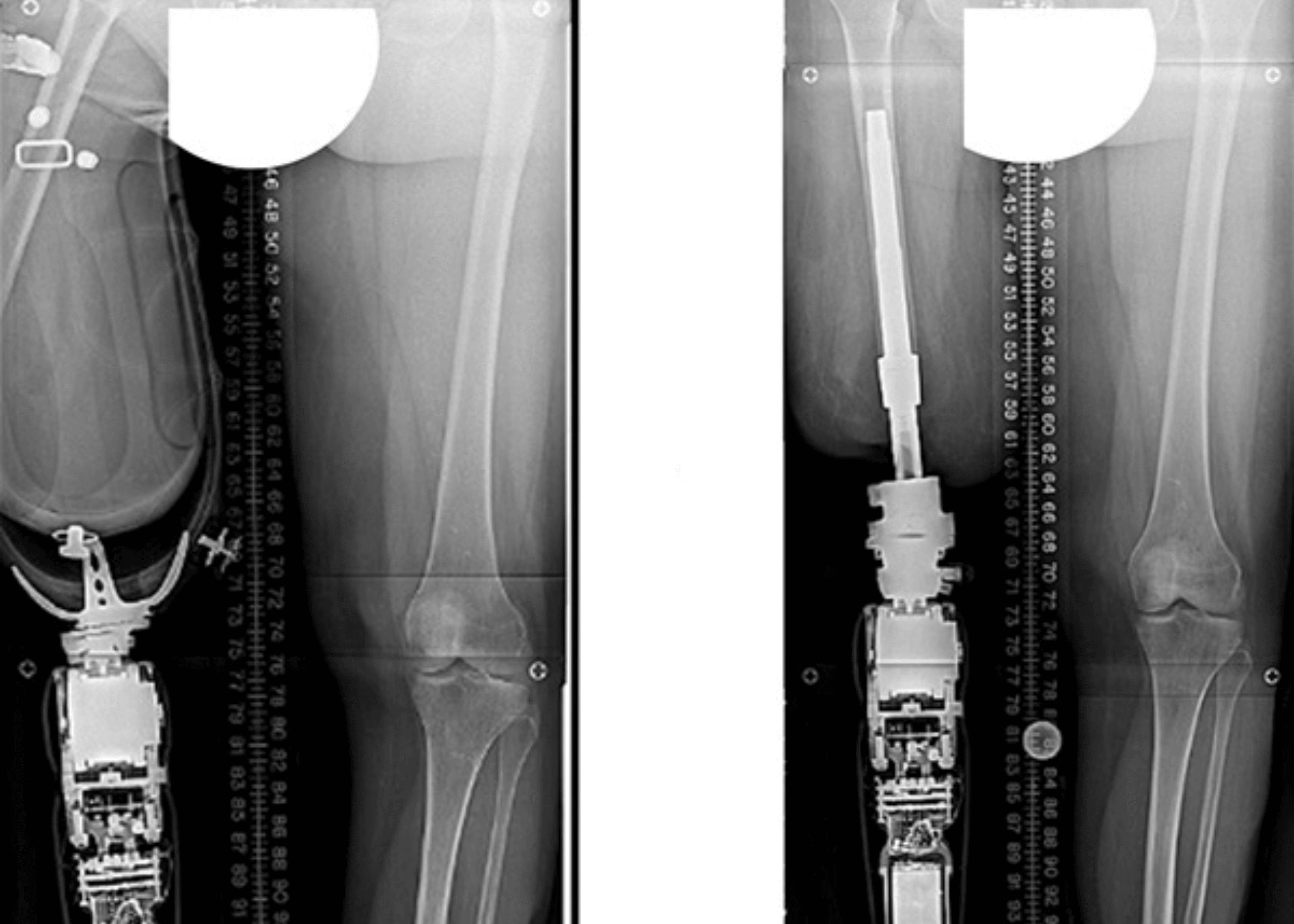
The patient presented with significant socket-related issues stemming from a short residual limb. Years of conventional prosthetic use had resulted in thickening of the distal stump and callosity formation, despite previous skin grafting interventions. As a non-smoker with Type 2 diabetes, the patient represented a suitable candidate for this advanced surgical approach.

Clinical Assessment

Examination revealed clear evidence of soft tissue thickening and callosity at the distal end of the residual limb, consistent with chronic socket-related pressure and friction. The medical team noted healed graft sites on distal sections of the stump without any signs of active infection or ulceration, confirming candidacy for the procedure.

Surgical Approach

Under the expert guidance of a specialized surgical team including Dr. Munjed Al Muderis, Dr. Michael George Uglow, Dr. Ahmed Hammouda, and anesthesiologist Dr. Ahmed Omran, the osseointegration procedure was meticulously performed at Burjeel Medical City’s state-of-the-art surgical facility.

The surgical technique involved:

  • Minimal skin incision using a corer to preserve tissue integrity
  • Conservative soft tissue dissection to maintain stability and vascularity
  • Preservation of posterior soft tissue and hyperkeratotic skin to minimize stoma infection risk
  • Sequential reaming of the distal tibial medullary canal from 8mm to 23mm
  • Fluoroscopy-guided broaching up to 24mm for precise implant positioning
  • Insertion of an OPL Tibial Type D implant (24×80mm) with press-fit fixation

This approach achieved excellent rotational stability with minimal bleeding upon tourniquet release, eliminating the need for flap refashioning.

Benefits of Osseointegration

Osseointegration represents a revolutionary advancement for amputees facing challenges with traditional socket prosthetics. By directly anchoring the prosthetic limb to the bone, this technique eliminates common issues such as:

  • Socket discomfort and pain
  • Skin irritation and breakdown
  • Limited range of motion
  • Difficulty with prosthetic fitting
  • Reduced proprioception and stability

Patients typically experience improved mobility, enhanced sensory feedback, and greater freedom from the limitations of socket-based systems.

Burjeel Medical City’s Advanced Surgical Capabilities

This procedure showcases Burjeel Medical City’s commitment to providing cutting-edge surgical interventions across its network of facilities in Abu Dhabi, Dubai, Al Ain, Sharjah, Al Dhafra, and Oman. With 70 operating rooms and specialized equipment including 7 catheterization laboratories, Burjeel Medical City’s surgical infrastructure supports complex procedures that transform patients’ lives.

Conclusion

The successful osseointegration procedure demonstrates the advanced surgical capabilities available at Burjeel Medical City facilities. By embracing innovative techniques, the team of orthopedic surgeons continues to provide life-changing solutions for patients with complex medical challenges, maintaining Burjeel Medical City’s position at the forefront of healthcare excellence in the region.

Experts

Prostate Cancer.jpg

Prostate Cancer – Symptoms, Causes & Treatment

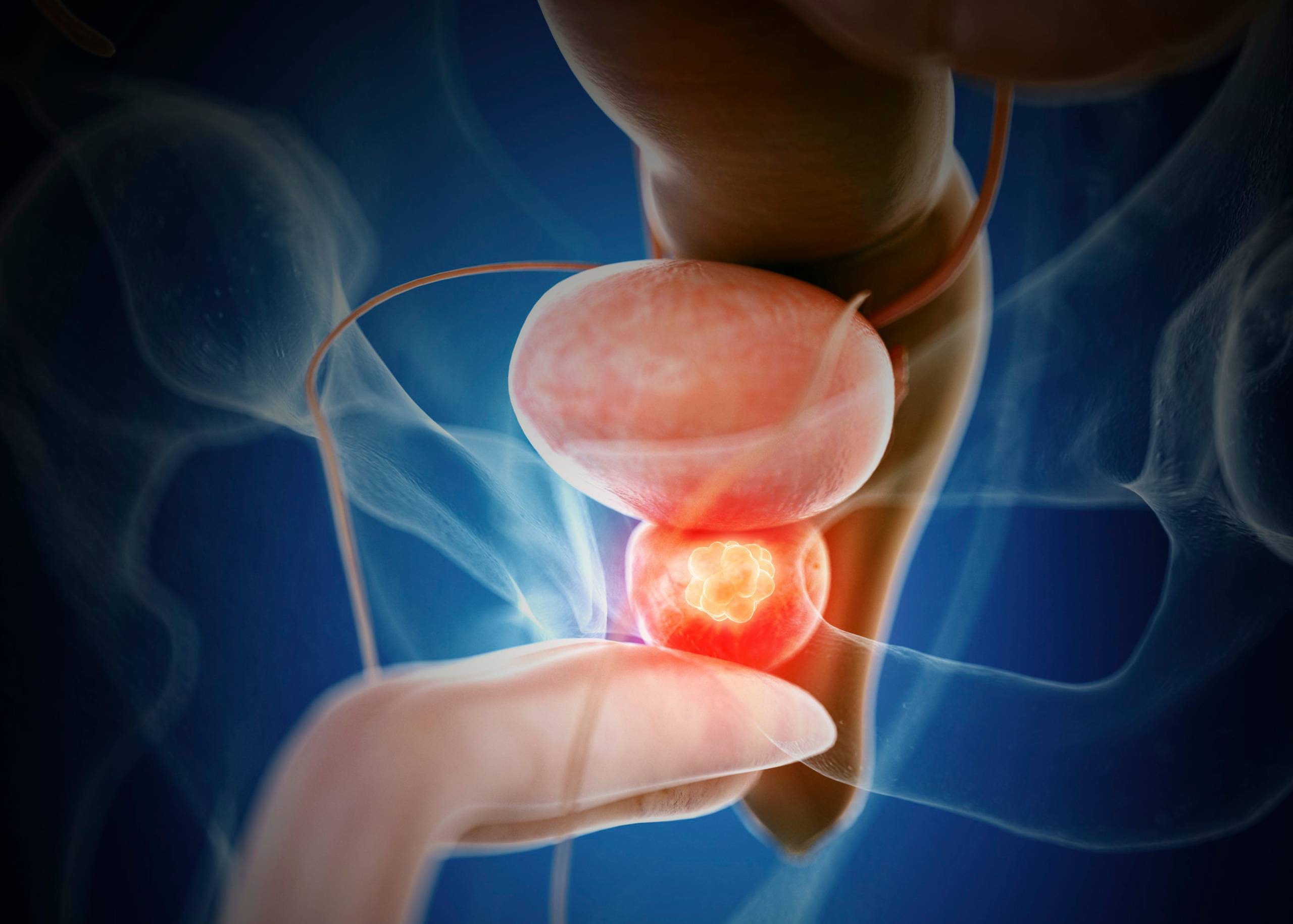
Prostate cancer is one of the most common cancers in men. The prostate is a gland that makes some of the fluid that helps to nourish and transport sperm. Prostate cancer starts when cells in the prostate grow abnormally and crowd out normal tissue. The tumor grows slowly at first, but as it gets bigger, it can damage nearby tissues, causing pain and other problems. Most prostate cancers grow slowly and do not spread to other parts of the body. But some are aggressive and have a high chance of spreading to other organs, such as the bones and lymph nodes.

Symptoms of Prostate Cancer

The most common signs are:

  • Difficulty urinating, either dribbling or a weak flow
  • A feeling that you have to urinate more often than usual
  • Pain or burning while urinating
  • Blood in your urine or semen

Causes of Prostate Cancer

It’s estimated that more than 40% of men will develop prostate cancer at some point. The causes of prostate cancer are not well understood, but researchers have found that certain factors can increase a man’s risk of developing it. These include

Age: As men get older, their risk of developing increases. It is more common in men over the age of 50.

Diet: Eating a diet high in red meat or dairy products may increase your risk.

Family History: If a close relative has been diagnosed with the condition, you’re at higher risk for developing it yourself.

Infection: Infection by the sexually transmitted virus HPV increases your risk of developing both low-grade and high-grade prostate cancers.

Diagnosis

Prostate cancer is diagnosed by a combination of physical examination, blood tests, and biopsies.

Physical Examination. The doctor will examine the prostate for swelling, lumps, or other abnormalities. The doctor may also order a digital rectal exam (DRE) to check the prostate for lumps and abnormalities.

Blood Tests Determine elevated levels of PSA (prostate-specific antigen) and DRE2-3 (a protein produced by many cancers).

Digital Rectal Exam (DRE) may also be performed to identify any abnormalities in the prostate.

Biopsy. The diagnosis of prostate cancer can be confirmed with a biopsy of tissue samples taken from your prostate gland during an examination known as a transrectal ultrasound guided needle biopsy (TRUS-NB). This procedure involves inserting an ultrasound probe into your rectum so that the doctor can see inside your body and guide a thin needle over your prostate gland.

Treatment

Treatment of prostate cancer varies based on the disease’s stage and the patient’s age. Treatment options include surgery, radiation therapy, hormone therapy, and chemotherapy. 

Radiation therapy is used for patients with early-stage disease that has not spread to local lymph nodes and for patients with advanced-stage disease that is confined to the prostate gland. In external beam radiation therapy (EBRT), a machine directs a beam of high-energy radiation at the tumor from outside the body. 

Brachytherapy involves placing radioactive seeds directly inside or around the tumor. 

Hormone therapy involves taking drugs such as luteinizing hormone-releasing hormone agonists (LHRH agonists) or antiandrogens to stop testosterone production in men who have advanced prostate cancer. The goal of hormonal treatment is to reduce levels of testosterone circulating in the bloodstream so that tumors do not grow as quickly as they would if testosterone levels were normal. 

Chemotherapy may be used alone or in combination with other treatments, such as surgery or radiation therapy, to treat advanced prostate cancer that has spread beyond its primary location (metastatic).

Surgery for Prostate Cancer

Surgery for prostate cancer involves removing the prostate gland or part of it. This surgery is called radical prostatectomy (RP). There are two types of RPs: open and robotic-assisted laparoscopic. The type of surgery you have depends on your tumor size and location, age, and other factors.

Radical prostatectomy is a surgical procedure to remove the prostate gland, including some surrounding tissue and lymph nodes. The surgeon makes an incision in the lower abdomen and removes the prostate gland through that opening. A catheter may be inserted into the urethra so urine can flow freely afterward. The surgeon then repairs any damage done during surgery, closes the incision, and stitches up any remaining skin openings on each side of the scrotum (in males).

At Burjeel Medical City, Abu Dhabi, we are dedicated to providing the best possible care for patients with prostate cancer. Our experienced team of uro-oncologists is committed to providing the highest quality of care and treatment for our patients. We understand the importance of providing comprehensive care for prostate cancer and are dedicated to providing the best possible outcomes for our patients. With our cutting-edge technology and expertise, we strive to provide the best possible care for our patients and ensure that they have the best chance.